Before an AI can judge a chest X-ray, it has to answer a humbler question first: where are the lungs? Get that boundary wrong and every diagnosis built on top of it inherits the mistake. This is the engine room that gets it right.
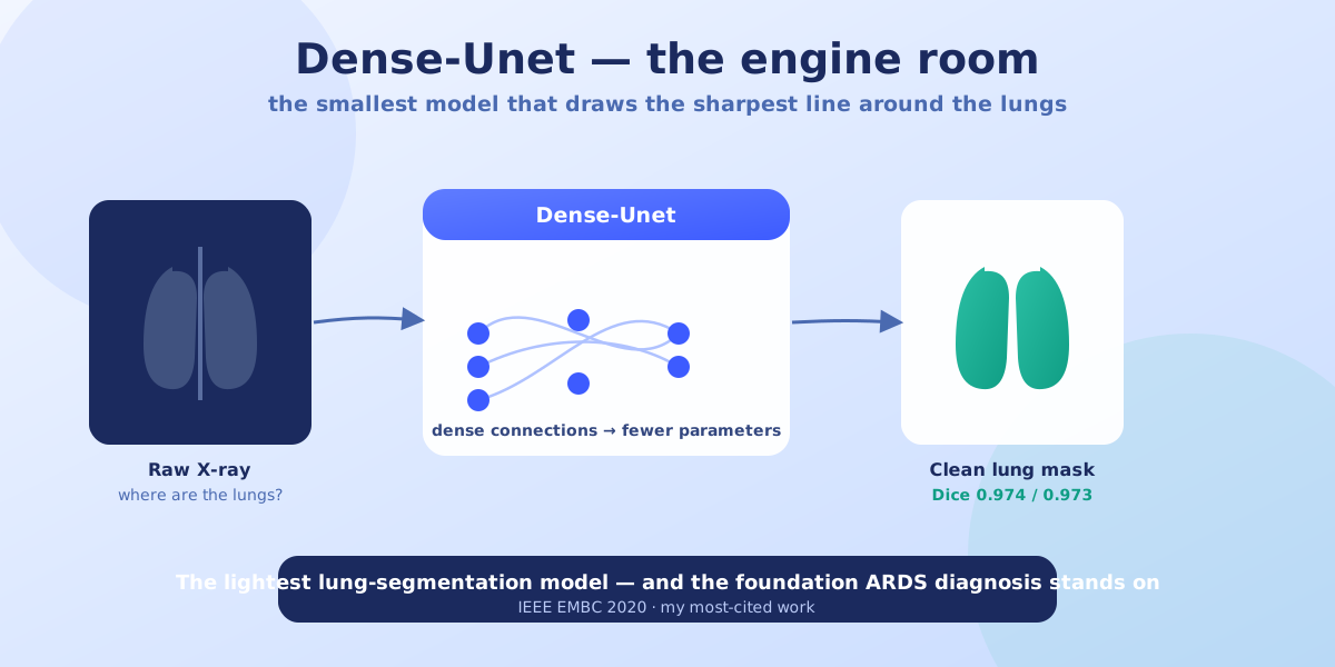
Dense-Unet is the model at its heart — and its headline is a paradox: it's both the most accurate and the lightest deep model proposed for segmenting lung fields in chest X-rays.
How it pulls that off. Big segmentation networks buy accuracy with millions of parameters — which eats memory and, worse, hurts how well the model handles X-rays it has never seen. Dense-Unet takes the opposite bet: dense connections wire information through the network so efficiently that it needs far fewer parameters to reach the same, or better, precision. Small model, sharp result, better generalization.
The result: a Dice score of 0.974 on the JSRT benchmark and 0.973 on Montgomery County — essentially tracing the lung boundary the way a careful human would, and fast enough to run in a clinical setting without a heavyweight server.
Why it exists. This isn't segmentation for its own sake. Precise, trustworthy lung boundaries are the foundation the whole pediatric-ARDS diagnostic system stands on — the clean canvas that makes the quadrant-by-quadrant judgment in my ARDS diagnosis project possible. Published at IEEE EMBC 2020, it became my most-cited work.
Under the hood (for the engineers): a U-Net variant with dense connectivity for maximal information flow at minimal parameter cost, tuned for the small, carefully-labeled datasets that real clinical work actually provides. Read it on PolyPublie.
Why it matters (for everyone else): the unglamorous, essential step — drawing the line around the lungs so precisely that everything downstream can be trusted.
Skills: Deep Learning · Digital Image Processing · Semantic Segmentation · Medical Imaging